How some brain cells hook up surprises researchers
November 3, 2010

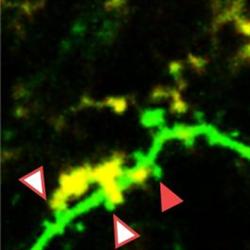
This two-photon image shows dynamic interaction between a microglial cell (yellow) and a neuron’s dendritic spines (green points) in the living brain. The spines with white arrows are being touched by the microglia; the red arrow points to a spine that had been touched by the microglia just five minutes earlier. (University of Rochester)
According to a recent study, immune cells known as microglia, long thought to be activated in the brain only when fighting infection or injury, are constantly active and likely play a central role in one of the most basic, central phenomena in the brain — the creation and elimination of synapses.
The finding, reported in the Nov. 2 issue of PloS Biology, means that the microglia cell may, in addition to its well-recognized duty of protecting the brain, also have direct involvement in creating the cellular networks at the core of brain behavior. Its apparent role as an architect of synapses comes as a surprise to researchers long accustomed to thinking of microglia as cells focused exclusively on keeping the brain safe from threats.
“When scientists talk about microglia, the talk is almost always about disease. Our work suggests that microglia may actively contribute to learning and memory in the healthy brain, which is something that no one expected,” said Ania Majewska, Ph.D., the neuroscientist at the University of Rochester Medical Center who led the work.
The group’s paper is a detailed look at how brain cells interact with each other and react to their environment swiftly, reaching out constantly to form new links or abolish connections.
First author Marie-Ève Tremblay, Ph.D., a post-doctoral associate in Majewska’s lab, used immunoelectron microscopy and two-photon microscopy to look at how microglia interact with synapses in the brains of healthy mice as their environment changed. In the experiments, the scientists looked into the brain while the mice were on a normal cycle of light and dark, while the mice were in the dark for several days, and again when the mice went back to a normal light/dark cycle.

Electron micrograph of a microglia cell (shown in black) touching surrounding cells in the synapse, including dendritic spines (pink) and axon terminals (blue). (University of Rochester)
The Rochester team found a high level of activity among microglia in response to the visual changes that the mice experienced. Even though scientists often say that microglia that are not actively fighting an injury or infection are “at rest,” scientists found that even under normal circumstances, microglia are still active.
“Why are microglia so dynamic? What are they contacting, and why, even when there is no injury?” asked Tremblay. “The idea that immune cells are always active in our brain, contributing to the ongoing process of learning and memory, really challenges current views of the brain.”
Most notably, the scientists found that microglia changed their activity in response to the environment. When the lights were off, microglia contacted more synapses, were more likely to reach toward a particular type of synapse, tended to be larger, and were more likely to appear to be poised to destroy a synapse. When the lights came back on, most of those activities reversed.
“The fact that microglia change their behavior based on visual input is a remarkable feat for a cell whose role supposedly is all about brain injury and disease. Just the fact that microglia can sense that something has changed in the environment is a novel idea,” said Majewska, who is assistant professor in the Department of Neurobiology and Anatomy.
The team showed how microglia constantly send out their extensions, which are like tentacles, often targeting synapses. In time-lapse video of their experiments, microglia dance across the screen, sending their extensions throughout the brain constantly. The cells also are known to travel remarkably quickly through the very dense and convoluted environment of the brain, traveling perhaps two millionths of a meter in a minute.
Tremblay and Majewska showed that microglia touch and wrap around synapses constantly and may have some say in deciding which synapses will survive and which will disappear. Microglia also appear key to creating or changing the extracellular space around synapses, a factor that would profoundly affect synapse function.
The team also found indications that microglia may be involved in destroying synapses through a process known as phagocytosis. Microglia had extensive interactions with tiny lollipop-shaped structures called dendritic spines, which are essential for a neuron’s ability to connect with other nerve cells that transmit excitatory signals. Eliminating dendritic spines is one way to destroy synapses.
The scientists found that dendritic spines that were touched by microglial processes during Tremblay’s first imaging session were more than three times as likely to be eliminated within the next two days compared to spines that were not. And microglia interacted more often with smaller dendritic spines, which generally indicate synapses that are either in the early stage of formation or that can easily be broken.
Years ago, brain signaling was thought to be the exclusive domain of neurons. During the last two decades, scientists have found that astrocytes also have vast signaling networks. Now, microglia also seem to be an important player in the brain’s ability to adapt immediately and constantly to the environment and to shift its resources accordingly.
The findings are timely for scientists who are increasingly studying links between the nervous and immune systems, Tremblay said. The role of microglial cells themselves is being looked at in an array of conditions, including Parkinson’s and Alzheimer’s diseases, schizophrenia, obsessive-compulsive disorder, and even autism.
Adapted from materials provided by at the University of Rochester Medical Center